
Seven Reasons Your Shoulder Pain Has Not Been Fully Explained
A patient walks in with shoulder pain that has been treated for a year. The MRI report says partial rotator cuff tear. There has been a corticosteroid injection, sometimes two. There has been physical therapy. There has been a recommendation for surgery or a recommendation to keep trying conservative care, depending on which physician was consulted last.
The pain is still there.
This is one of the most common presentations in regenerative musculoskeletal medicine, and it is almost never a story about a single structure. It is a story about an incomplete evaluation, an imaging report that was treated as the diagnosis, and a treatment plan that was built on top of that mistake.
The shoulder is not a structure. It is a region. It includes the glenohumeral joint, the acromioclavicular joint, the four rotator cuff tendons, the long head of the biceps tendon, the joint capsule, the shoulder blade (scapula), the cervical spine, the upper ribs, and the fascial and muscular system that coordinates them. Any one of these can generate pain. Multiple can do so simultaneously. The cervical spine can refer pain into the shoulder in a pattern that is clinically indistinguishable from local shoulder pathology.
When the evaluation stops at the MRI report, the diagnosis stops at the first identifiable abnormality on the scan. That is not the same thing as identifying what is actually causing the patient’s pain.
What Imaging Cannot Tell You
Imaging shows anatomy. It does not show how the shoulder behaves under load.
It does not show how the scapula rotates during arm elevation, whether the rotator cuff activates appropriately to center the humeral head, whether the biceps tendon is being repeatedly stressed during reaching, whether the posterior capsule has become restricted, whether cervical nerve roots are referring symptoms into the deltoid and arm, or whether the patient compensates around a finding the radiologist correctly identified but that may not be the primary driver of pain.
A partial rotator cuff tear on MRI may be the diagnosis. It may also be coincidental anatomy in a shoulder whose pain is being driven by a different structure entirely. The MRI cannot tell the difference. The examination can.
Seven Sources of Shoulder Pain That Are Commonly Missed
1. Rotator cuff pathology that the tear report does not describe
The rotator cuff is four tendons that center the humeral head during all shoulder motion. Pain can arise from tendinopathy, calcific deposits, bursal irritation, inadequate cuff activation, or chronic overload from poor scapular mechanics. None of these are “tears.” All of them can produce the same pain pattern. When an MRI report identifies a partial tear and the conversation stops there, the functional contributors to cuff stress, the actual reasons the cuff is irritated, go unaddressed. The tear becomes the explanation. The mechanics that produced it remain active.
2. AC joint degeneration that nobody is asking about
The acromioclavicular joint is small. It is also one of the most reliable hidden pain generators in the shoulder. It produces pain with cross-body reaching, with bench press and pushup movements, with golf and tennis follow-through, and with sleeping on the affected side. It is anatomically distinct from the subacromial space, which means an injection placed under the acromion does nothing to address AC joint pathology even when delivered with technical precision.
Patients who have received one or two corticosteroid injections without relief are frequently told the injection failed. The injection did not fail. It treated the wrong structure.
3. Biceps tendon involvement that the standard exam misses
The long head of the biceps tendon runs through the bicipital groove at the front of the shoulder. It can become primarily inflamed, secondarily irritated by adjacent rotator cuff pathology, or involved in superior labral pathology. Patients describe pain with lifting, carrying, pulling, reaching into the back seat, and supinating the forearm against resistance.
A shoulder examination that focuses on overhead motion and resisted abduction will miss biceps tendon involvement. A complete examination includes specific provocative testing of the tendon in the groove and tests the tendon’s behavior during the specific motions the patient identifies as painful.
4. Glenohumeral joint arthritis presenting as rotator cuff pain
Glenohumeral arthritis can be subtle in its early stages. Patients maintain functional overhead motion. They lose rotation. They develop morning stiffness. They feel grinding. They have difficulty reaching behind their back. None of these features are required to be present, and the symptom complex frequently overlaps with rotator cuff and bursal patterns.
When the joint is the source, the treatment changes. Intra-articular intervention is anatomically and clinically different from subacromial or peri-tendinous intervention. Identifying glenohumeral arthritis early, before it dictates the entire conversation, is part of a complete shoulder evaluation.
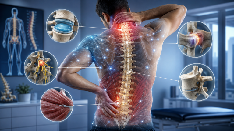
5. Cervical referral that the patient does not recognize as a neck problem
The cervical spine refers pain into the shoulder, the upper arm, the periscapular region, the elbow, the forearm, and the hand. Patients frequently describe the shoulder as the location of their pain because that is where they feel it most. They do not have neck pain. They do not have hand symptoms. The shoulder is where it hurts.
Cervical pathology can produce a symptom complex that overlaps almost perfectly with rotator cuff and subacromial pain. A cervical screen, including range of motion, neurological assessment, and provocative testing for nerve root involvement, is a non-optional component of any shoulder evaluation. When it is skipped, patients who have neck-driven shoulder pain receive shoulder injections that cannot help them, sometimes for years.
6. Posterior capsule tightness that imaging will not show
The posterior capsule controls internal rotation and contributes to the centered position of the humeral head during elevation. When it becomes contracted, the humeral head translates anteriorly and superiorly during arm motion, producing impingement, increased cuff stress, and biceps tendon loading. This is a functional finding. The MRI may be unremarkable. The pathology is in the mechanics, not the static anatomy.
Posterior capsule tightness is identified on examination. It is treated with targeted mobilization and rehabilitation, not with injection. When it is present and not addressed, every cuff-directed and bursa-directed intervention fights against an unrecognized mechanical fault that continues to load the structures the injection is trying to quiet.
7. Scapular dyskinesis as a cause, not a consequence
The scapula is the moving platform on which the rotator cuff operates. When scapular mechanics are impaired, the cuff is forced to work against a poorly positioned and poorly controlled base. The biceps tendon is stressed. The AC joint is compressed. Cervical and periscapular musculature become hypertonic. The shoulder hurts in patterns that look like multiple structural problems, because the system is failing in multiple locations at once.
Scapular dyskinesis is often identified after the fact and attributed to the patient’s pain. In a significant subset of patients, particularly those with chronic, recurrent, or treatment-resistant shoulder pain, the dyskinesis is the upstream cause of the entire pain pattern. Treating the downstream structures while the scapular mechanics remain unaddressed is the clinical equivalent of mopping the floor while the pipe leaks.
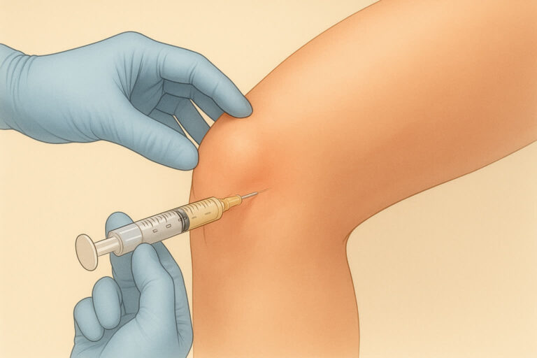
Why Injection Accuracy in the Shoulder Is Not a Technical Footnote
The shoulder contains multiple injection targets in close anatomical proximity. The subacromial space, the AC joint, the glenohumeral joint, the biceps tendon sheath, and individual rotator cuff tendons are not interchangeable. An injection placed accurately into the subacromial space has no effect on AC joint pathology. An injection placed into the glenohumeral joint does not treat biceps tendinopathy. Each target requires its own placement.
Landmark-based shoulder injections, which estimate placement from surface anatomy without imaging confirmation, have documented accuracy limitations that are particularly pronounced for the AC joint and biceps tendon sheath. The research is consistent. When the target is small and the anatomical neighborhood is dense, accurate placement requires real-time imaging confirmation.
At Precision Regenerative Medicine™, every shoulder injection is performed under ultrasound guidance. Not because guidance is a marketing feature. Because without it, the procedure becomes an estimate.
What a Complete Shoulder Evaluation Requires
I am Dr. Tammy J. Penhollow, DO. I am dual board-certified in Anesthesiology and Pain Medicine, Stanford fellowship-trained, and the founder of Precision Regenerative Medicine™ in Scottsdale, Arizona. I evaluate shoulders by examining the region, not just the structure named in the MRI report. I do not perform blind injections. I do not use corticosteroids in my practice.
A complete shoulder evaluation at Precision Regenerative Medicine™ includes:
- Detailed pain history with attention to position, load, and motion triggers that point toward specific structural contributors
- Prior treatment review, including what was injected, where, how it was guided, what relief was obtained, and how that relief evolved with repeat treatment
- Physical examination of the rotator cuff, biceps tendon, AC joint, and glenohumeral joint with structure-specific provocative testing
- Active and passive range of motion assessment, with specific attention to rotational asymmetry that suggests capsular involvement
- Cervical spine screen, including neurological assessment and provocative testing for radicular contribution
- Scapular mechanics evaluation during arm elevation and resisted motion
- Imaging correlation, in which MRI findings are interpreted alongside examination and symptom behavior rather than treated as the diagnostic answer
- Metabolic and systemic assessment relevant to tissue healing capacity if regenerative intervention is being considered
How Treatment Changes When the Diagnosis Is Complete
When the actual pain generator is correctly identified, the treatment plan often looks quite different from what would have been proposed based on the imaging alone.
Rotator cuff tendinopathy that is being driven by scapular dyskinesis is treated by addressing scapular mechanics first. Biceps tendon pain is treated at the biceps tendon. AC joint degeneration is treated at the AC joint. Glenohumeral arthritis is treated intra-articularly. Cervical referral is treated at the cervical spine and may not require shoulder intervention at all. Posterior capsule tightness is treated with targeted mobilization. Each structure gets the intervention it actually needs.
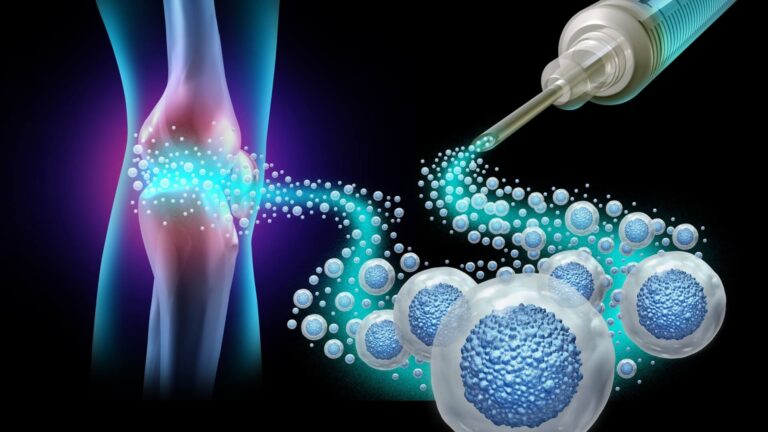
When regenerative intervention is appropriate, options at Precision Regenerative Medicine™ include image-guided PRP, bone marrow aspirate concentrate, and prolotherapy directed at confirmed tissue targets including the rotator cuff tendons, the biceps tendon sheath, the glenohumeral joint, and the AC joint. Procedures are autologous. Every injection is ultrasound-guided. Rehabilitation and movement correction are sequenced with the biology, because biology placed accurately into a mechanically dysfunctional shoulder will fight against the mechanics every day it is healing.
This integrated approach is delivered through the Joint Boost System™, the clinical framework I developed for treating major joints as coordinated mechanical and biological units. Joint Boost System™ addresses scapular mechanics, cervical contribution, capsular mobility, and neuromuscular control alongside any biologic intervention at the shoulder. The same framework applies across the knee, hip, and elbow, with joint-specific adaptations. The biology and the mechanics are sequenced together, not delivered separately.
Frequently Asked Questions
Why does shoulder pain persist after multiple injections?
Persistent shoulder pain after multiple injections almost always indicates an incomplete diagnosis. The injections may have been technically appropriate for the structure named in the imaging. They cannot help if that structure is not the actual driver of pain. The next step is not another injection. It is a complete regional evaluation.
Can shoulder pain come from the neck?
Yes, frequently. Cervical pathology refers pain into the shoulder in a pattern that can be clinically indistinguishable from local shoulder pathology. A cervical screen is a required component of any shoulder evaluation. When it is skipped, neck-driven shoulder pain receives shoulder treatment that cannot resolve it.
Is PRP appropriate for shoulder pain?
PRP may be appropriate for selected shoulder tendon, joint, and soft tissue pain patterns once a complete evaluation has confirmed the actual pain generator and the tissue picture supports biologic intervention. PRP is not a default treatment for all shoulder pain. The diagnosis determines whether it is indicated, where it should be delivered, and what should be done alongside it.
What is scapular dyskinesis and why does it matter clinically?
Scapular dyskinesis refers to altered scapular position or motion during arm movement. Because the scapula is the platform on which the rotator cuff operates, poor scapular control increases stress on the cuff, biceps tendon, and AC joint. In patients with chronic or recurrent shoulder pain, scapular dyskinesis is frequently the upstream mechanical driver of the entire pain pattern, not a secondary finding.
What does image guidance actually change in a shoulder injection?
Image guidance allows direct visualization of the target structure, the needle, and the delivery of the injectate. Without it, placement is estimated from surface landmarks. For small, closely spaced shoulder targets like the AC joint and biceps tendon sheath, the accuracy difference is clinically significant. An injection that misses its intended target produces no therapeutic effect, even when the technique appears competent.
The Diagnosis Is the Work
Patients with shoulder pain that has not resolved despite multiple treatment attempts do not need another injection delivered with more conviction. They need someone to evaluate the entire shoulder region, identify the actual structures contributing to their pain, screen for cervical referral, examine the scapula, test the posterior capsule, and build a treatment plan that addresses what is actually happening.
That is the work that prevents the next round of unsuccessful treatments. It is the work that determines whether a regenerative intervention will produce a result or simply join the list of things that have already been tried. At Precision Regenerative Medicine™ in Scottsdale, Arizona, that evaluation is the starting point for every shoulder patient, before any treatment is discussed.
To request a consultation, complete the contact form at precisionmedprp.com/contactus/ and Dr. Penhollow’s office will follow up with you directly.